


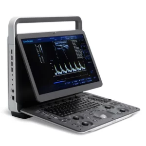
Ultrasound Color Doppler SonoScape P20
Original price was: 249999,00 kr.199999,00 krCurrent price is: 199999,00 kr.
exklusvie moms.SPECS
P20 system enhances flexibility and intelligence to a new level. Stable as ever, P20 improves its signal transmission and reception processors which leads to higher sensitivity and more accurate echo detection. What’s more, P20 is equipped with a wide range of transducers which adopt innovative technologies and therefore promises a confident diagnostic experience. Consistent quality performance across applications.
Enhanced with advanced imaging architecture and probe technologies, the system enlarges diagnostic capacity and quality across a wide range of applications
- μ-Scan
- Adaptive Multi-beam Imaging
- Dynamic Color
- Single Crystal Transducer
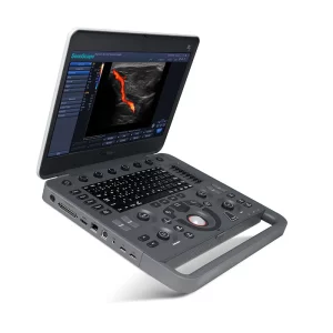
Ergonomic Design
- 21,5” LED monitor with articulated arm
- 13,3” Adjustable touch screen
- Rotatable and height adjustable control panel
Available with a range of high definition probes:
- Linear Array L741 (Vascular, Small parts, MSK etc.), 4-16 MHz/ 46 mm
- Linear Array L742(Vascular, Small parts, MSK etc.), 4-16 MHz/ 38 mm
- Linear Array L752(Vascular, Small parts, MSK etc.), 4-16 MHz/ 52 mm
- Linear Array 12L-A(Vascular, Small parts, MSK etc.), 3-17 MHz/ 52 mm
- Linear Array 12L-B(Vascular, Small parts, MSK etc.), 3-17 MHz/ 38 mm
- Convex Array 3C-A (Abdominal, Obstetrics, Gynecology), 1.0-7.0 MHz/ R50 mm
- Convex Array C1-6 (Single crystal) (Abdominal, Obstetrics, Gynecology), 1.0-8.0 MHz/ R50 mm
- Convex Array C613(Cardiology, Pediatrics), 3-15 MHz/ R14mm
- Phased Array 3P-A (Cardiac, Transcranial), 1.0-5.4 MHz
- Phased Array 7P-B (Cardiac, Transcranial, Infant), 2-9MHz
- Phased array S1-5 (Single crystal) (Cardiac, Transcranial), 1.0-7.0 MHz
- Endocavity 6V3 (Gynecology, Obstetrics, Urology), 3-15 MHz/ R10 mm
- Endocavity 6V1 (Gynecology, Obstetrics, Urology), 3-15 MHz/ R11 mm
- Endocavity 6V3A (Gynecology, Obstetrics), 3-15 MHz/ R8 mm
- Endocavity 6V7 (Gynecology, Obstetrics), 3-15 MHz/ R10 mm
- Volumetric Convex Array VC6-2(Obstetrics, Abdominal, Gynecology), 2-6.8 MHz/ R40 mm
- Volumetric Endocavity VE9-5 (Obstetrics, Gynecology, Urology), 2-13 MHz/ R10 mm
- Biplane BCL10-5 (Urology), Convex 3.9-11 MHz/ R10mm, Linear 6-15 MHz/ 60mm
- Phased Array Transesophageal MPTEE (Cardiac), 4-13 MHz
- Linear Array 10I2 (Intra-operative), 4-16 MHz/ 25mm
- Linear Array 12LI-A (Intra-operative), 4-16MHz/ 33mm
- Linear Array 12LT-A (Intra-operative), 4-16MHz/ 33mm
- Convex Array 6CT-A (Intra-operative), 3-15MHz/ R40mm
- Convex Array 6CI-A (Intra-operative), 3-15MHz/ R40mm
P20 Standard Configurations:
- P20 Main Unit size: 751×518×1350mm
- 21.5″ High Resolution LED Color Monitor
- 13.3″ High Resolution Touch Screen
- Height Adjustable and Rotatable Operation Panel
- Extended Keyboard
- Five Transducer Connectors (Four Active + One Parking)
- One Pencil Transducer Connector
- Wifi Module
- Hard Disk 500G
Optional configurations:
- The Fifth Active Transducer Connector
- ECG (Hardware / Cables: Must be Configured with ECG Software)
- Built-in Battery
- Built-in DVD
P20 Standard Configurations:
- μ-Scan (2D Speckle Reduction Technology)
- B (2B & 4B) Mode
- Color Doppler Flow Imaging
- Pulse Wave Doppler Imaging
- HPRF
- Continuous Wave Doppler Imaging
- Power Doppler Imaging / Directional Power Doppler Imaging
- M Mode
- Tissue Harmonic Imaging
- Tissue Specific Imaging
- Simult Mode (Triplex)
- Pulse Inversion Harmonic Imaging
- Spatial Compound Imaging
- LGC (Lateral Gain Compensation)
- Full Screen Zoom
- Image Rotation
- Real-time 2D Panoramic Imaging
- Biopsy Guide
- PW Auto Calculation
- Auto NT
- Auto IMT
- Auto EF
- TEI Index
- S-Guide
- Auto Optimization (One Button Optimization for 2D / Color / PW)
- DICOM 3.0: Store / C-Store / Worklist / MPPS / Print / SR / Q&R
Optional Configurations:
- ECG (Configured with ECG Hardware and Cables)
- Color M Mode
- Anatomic M Mode
- Tissues Doppler Imaging
- Trapezoidal Imaging
- Real-time Color Panoramic Imaging
- VIS-Needle (Needle Visualization Enhancement)
- Freehand 3D Imaging
- Static 3D / 4D
- Auto Face (Configured with 3D / 4D Module at the Same Time)
- S-Live (S-Live / S-Live Silhouette)
- S-Depth
- AVC Follicle (Auto Volume Calculation)
- Stress Echo
- C-xlasto (Compression Elastography Imaging)
- Contrast Imaging
Applicaitions:
- Abdomen Package
- Gynecology Package
- Obstetrics Package
- Cardiology Package
- Small parts Package
- Urology Package
- Vascular Package
- Pediatrics Package
Value without Compromise, Treat with Confidence
 C-Xlasto Imaging

With C-xlasto Imaging, P20 enables comprehensive quantitative elastic analysis. Meanwhile, C-xlasto on P20 is supported by linear, convex and transvaginal probes, to ensure good reproducibility and highly consistent quantitative elastic results.
 Contrast Imaging

Contrast Imaging with 8 TIC curves allows doctors to assess perfusion dynamics in a wide range of clinical settings, including both the location and evaluation of lesion parts.
 S-Live
S-Live allows for detailed visualization of subtle anatomical features, thereby enabling intuitive diagnosis with real-time 3D images and enriching patient communication.
 Pelvic Floor 4D Transperineal 4D pelvic floor ultrasound can provide useful clinical values in assessing the vaginal delivery impact on the female anterior compartment, judging whether the pelvic organs are prolapsed or not and the extent, determining if the pelvic muscles were torn accurately.
 Anatomic M Mode
Anatomic M Mode helps you observe the myocardial motion at different phases by freely placing sample lines. It accurately measures the myocardial thickness and the heart size of even difficult patients and supports the myocardial function and LV wall-motion assessment.
 Tissue Doppler Imaging

P20 is endowed with Tissue Doppler Imaging which provides velocities and other clinical information on myocardial functions, facilitating clinical doctors with the ability to analyze and compare the motions of different parts of the patient’s heart.
Youtube:
Related products
-
I fokus
B/W Ultrasound SonoScape E1 With 1 Linear Probe L746
Original price was: 74999,00 kr.49999,00 krCurrent price is: 49999,00 kr. Add to cart -
I fokus
Ultrasound Color Doppler SonoScape P20 With 2 Probe
Original price was: 259999,00 kr.189999,00 krCurrent price is: 189999,00 kr. Add to cart

Reviews
There are no reviews yet.